Digital X-Ray Services
Digital X-Ray provides quick imaging of bones, chest, and dental structures-commonly used for trauma, pulmonary, and skeletal evaluations.
What is an X-Ray?
X-Ray imaging uses ionizing radiation to create images of internal structures. Modern digital X-Ray captures high-quality images at low radiation doses and is extremely fast, making it ideal for emergency and routine diagnostic use.
Common X-Ray Exams
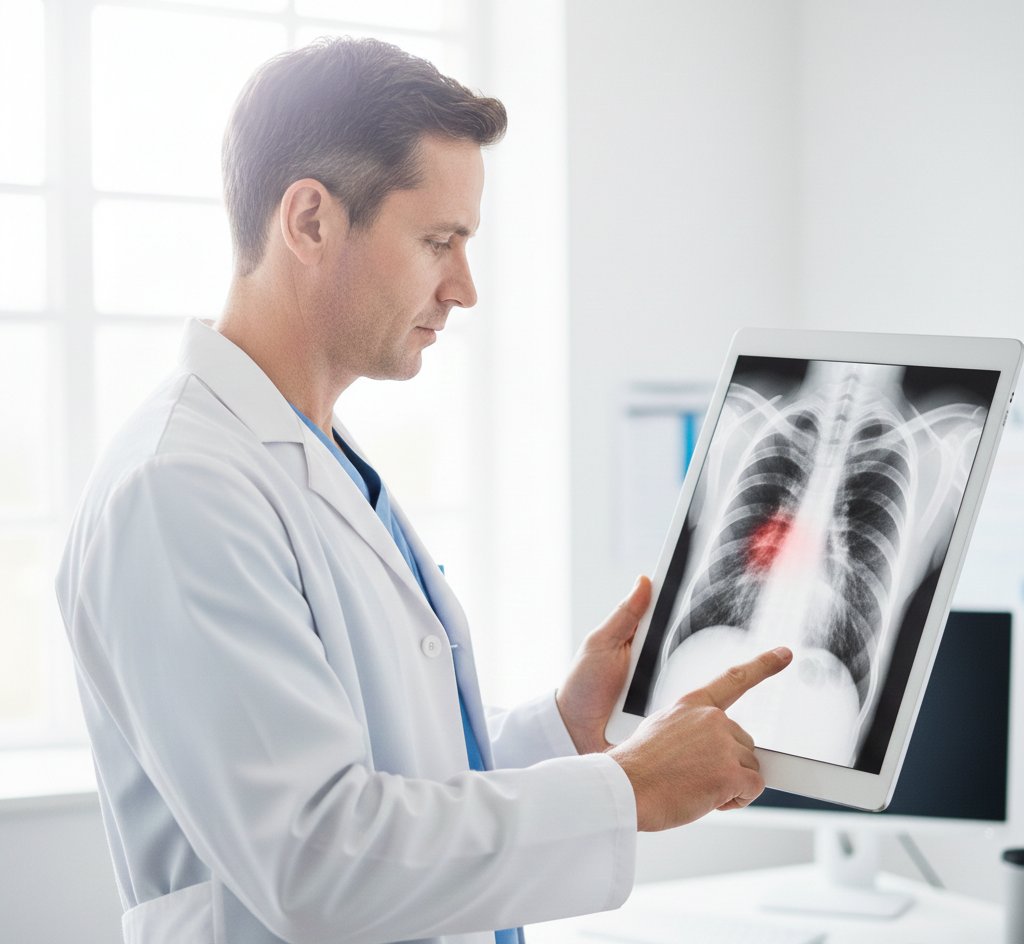
- Chest X-Ray - evaluate the lungs, heart, and chest wall; used to detect pneumonia, heart failure, or lung nodules.
- Musculoskeletal (Skeletal) X-Ray - assess bones and joints for fractures, dislocations, degenerative changes (arthritis), or bone tumors.
- Spine Imaging (Cervical, Thoracic, Lumbar) - visualize the vertebrae to diagnose alignment issues, disc degeneration, or spinal trauma.
- Abdominal X-Ray (KUB) - examine the kidneys, ureters, and bladder; often used to detect kidney stones or intestinal obstructions.
- Pelvic & Hip Imaging - evaluate hip joint integrity, pelvic fractures, or pre-operative planning for joint replacement.
- Sinus & Skull X-Ray - assess for sinusitis, facial fractures, or abnormalities in the cranial structure.
- Soft Tissue X-Ray - identify foreign bodies (such as glass or metal) or unusual swelling in the extremities.
- Pediatric X-Ray - specialized low-dose imaging tailored for children, including scoliosis series and bone age studies.
Patient Preparation
- Remove jewelry, watches, or metal objects near the area to be imaged.
- Pregnancy: inform the technologist if you are pregnant or suspect pregnancy.
- No fasting is typically required.
Reports & Turnaround
Our radiologists review X-Ray exams and final reports are typically sent to the referring physician within 48 hours. Urgent findings are communicated immediately.
Schedule an Appointment
