PET - Positron Emission Tomography
PET provides metabolic and physiologic imaging using radiotracers to detect disease at the molecular level.
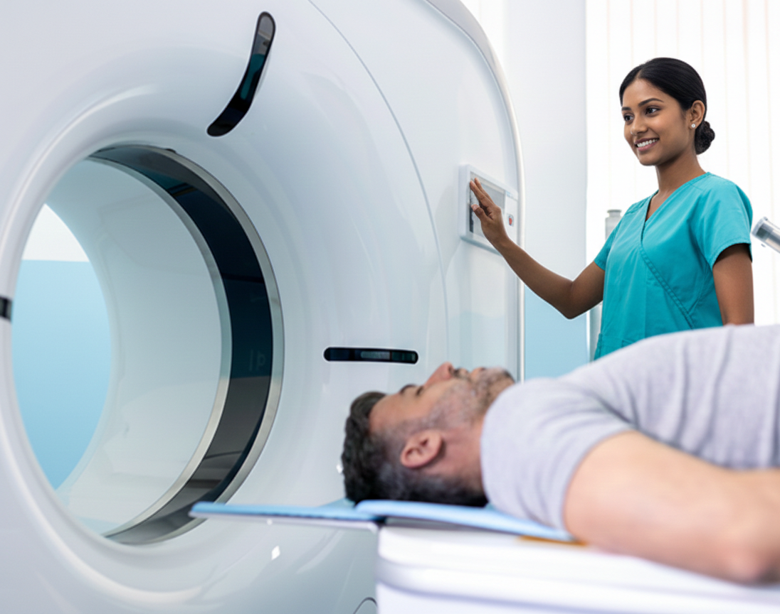
What is PET?
Positron Emission Tomography (PET) uses radiotracers that emit positrons. When combined with CT or MRI, PET provides highly sensitive functional imaging for oncology, cardiology, and neurology.
Common PET Exams
PET imaging is tailored to specific clinical questions and diseases. Below are our primary imaging domains:
Oncology (Cancer Imaging)
PET/CT is the "gold standard" for metabolic and functional imaging to detect cancer and monitor treatment response.
Diseases Evaluated:
- Breast, lung, and thyroid cancers
- Lymphoma
- Colorectal cancer
- Prostate cancer
Protocols:
- FDG-PET: Uses a glucose-like tracer to identify rapidly growing cancer cells—the most common oncology scan.
- PSMA PET: Specifically used to detect very small clusters of prostate cancer that are invisible on standard scans.
- Staging & Restaging: Determining if cancer has spread (metastasis) or returned after treatment (recurrence).
Neurology (Brain Imaging)
Used to examine the brain for specific disorders and functional activity patterns.
Diseases Evaluated:
- Alzheimer's disease and other forms of dementia
- Parkinson's disease
- Epilepsy (seizure disorders)
Protocols:
- Memory Clinic Evaluation: Specialized care for memory loss supported by advanced PET imaging.
- Brain Metabolism Scans: Mapping glucose usage patterns to pinpoint areas of dysfunction or seizure foci.
- Amyloid & Tau PET: Detecting protein abnormalities associated with neurodegenerative diseases.
Cardiology (Heart Imaging)
Evaluates organ function and blood flow without invasive procedures, providing critical information for cardiac risk stratification and treatment planning.
Diseases Evaluated:
- Coronary artery disease
- Heart attack damage (myocardial infarction)
- Myocardial inflammation and cardiomyopathies
Protocols:
- Perfusion & Viability Scans: Checking blood flow to the heart muscle and identifying healthy vs. damaged tissue (includes Cleerly® and HeartFlow® PET-based workflows as clinically indicated).
- Hibernating Myocardium Assessment: Determining if heart muscle would benefit from procedures like angioplasty or bypass surgery.
- Rubidium (Rb-82) Perfusion PET: Dynamic perfusion imaging for stress and rest assessment of myocardial blood flow.
- FDG Viability PET: Identifying viable but dysfunctional myocardium that may recover after revascularization.
Other PET Tracers
Beyond the primary domains above, we offer specialized PET tracers including amyloid PET, Ga-68, and other tracer studies when ordered by the referring physician for specific clinical indications.
Disclaimer: This information is for educational purposes only. Please consult a qualified medical professional for diagnosis or treatment recommendations.
Patient Preparation
- Bring prior imaging and the referring provider's order.
- Follow specific fasting or medication instructions provided at scheduling (varies by study).
- Hydration is typically encouraged unless instructed otherwise.
- Inform us if you are pregnant or breastfeeding.
Reports & Turnaround
Exams are interpreted by our board-certified physicians. Routine reports are delivered to the referring provider within 48 hours; urgent findings are communicated immediately.
Schedule an Appointment
