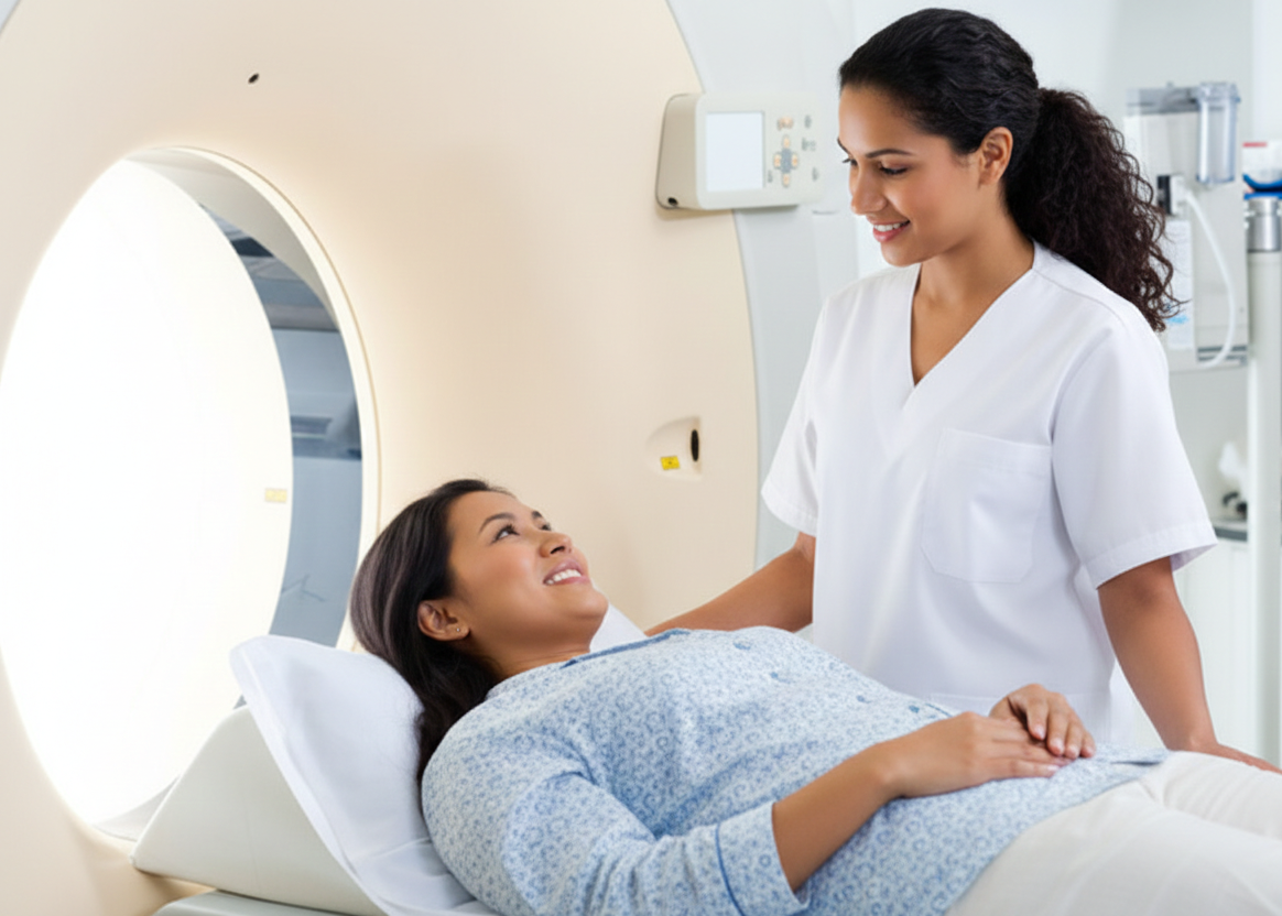
Nuclear Medicine
Nuclear medicine uses small amounts of radioactive tracers to evaluate organ function and physiology. It provides unique diagnostic information not available with conventional imaging.
What is Nuclear Medicine?
Nuclear medicine involves administering tiny amounts of radioactive tracers that accumulate in specific organs or tissues. A gamma camera detects the tracer distribution to produce images that reflect physiological function, such as thyroid uptake, bone turnover, or renal clearance.
Common Nuclear Medicine Exams
Nuclear medicine imaging uses radiopharmaceuticals to assess organ function and detect disease at the molecular level. Below are our primary nuclear medicine protocols:
Bone Scan
This protocol is the most effective for detecting changes in bone metabolism long before they appear on a standard X-ray.
Diseases Diagnosed:
- Metastatic bone cancer (spreading from breast, prostate, or lung)
- Osteomyelitis (bone infection)
- Stress fractures
- Paget's disease
- Arthritis
Protocol: Uses a radiopharmaceutical (99mTc-MDP) to identify areas of increased bone turnover.
Thyroid Uptake & Scan
Used to evaluate the health and function of the thyroid gland in the neck.
Diseases Diagnosed:
- Hyperthyroidism (overactive thyroid)
- Hypothyroidism (underactive thyroid)
- Graves' disease
- Hashimoto's thyroiditis
- Thyroid nodules or cancer
Protocol: Involves swallowing a small amount of radioactive iodine to see how the gland absorbs it.
Hepatobiliary (HIDA) Scan
A specialized scan to check the liver, gallbladder, and bile ducts for structural and functional abnormalities.
Diseases Diagnosed:
- Acute cholecystitis (gallbladder inflammation)
- Bile duct blockages
- Biliary atresia in infants
- Postoperative bile leaks
Protocol: Tracks the flow of bile from the liver into the small intestine.
Renal (Kidney) Scan
Measures kidney performance and structural health, functional output, and drainage patterns.
Diseases Diagnosed:
- Renovascular hypertension (high blood pressure from kidney issues)
- Renal artery stenosis
- Obstructions in the ureters
- Monitoring of kidney transplants
Protocol: Often used as a safe alternative for patients with allergies to CT or MRI contrast.
Additional Key Protocols
Specialized nuclear medicine studies available:
- Lung Scan (V/Q): Primarily used to detect Pulmonary Embolism (PE) or evaluate lung function before surgery.
- Gastric Emptying Study: The "gold standard" for diagnosing gastroparesis (slow stomach emptying) or rapid emptying syndrome.
- Myocardial Perfusion Scan: Evaluates blood flow to the heart muscle to identify coronary artery disease and heart attack damage.
- MUGA Scan: Radionuclide study for accurate left ventricular ejection fraction (LVEF) assessment, pre/post chemotherapy and cardiology evaluation.
- Parathyroid / Thyroid Therapy: Parathyroid localization studies and therapeutic options including I-123/I-131 whole-body scans, Thyrogen administration, and post-I-131 therapy imaging when requested.
- SPECT / Thallium / Technetium Studies: Nuclear cardiology and infection imaging using SPECT, Thallium, or Technetium-based tracers for infection/Gallium imaging.
Disclaimer: This information is for educational purposes only. Please consult a qualified medical professional for diagnosis or treatment recommendations.
Patient Preparation
- Bring prior imaging and the referring provider's order.
- Some studies require withholding certain medications (e.g., thyroid medications) - specific instructions will be provided when scheduling.
- Hydration is often encouraged; follow fasting instructions when indicated for specific exams.
- Inform us if you are pregnant or breastfeeding; tracer-specific precautions may apply.
Reports & Turnaround
Exams are interpreted by our board-certified nuclear medicine physicians and radiologists. Final reports are sent to the referring physician within 48 hours. Urgent or unexpected findings are communicated immediately.
Schedule an Appointment
